Fighting oral cancer with bioengineered chewing gum
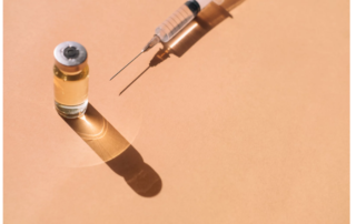
Written by: Deborah Stull Research led by Penn Dental’s Henry Daniell shows that antiviral and antibacterial chewing gums reduce the levels of three microbes linked to worse outcomes in oral cancers, paving the way for more effective and affordable therapies. esearchers led by Henry Daniell of the School of Dental Medicine have shown that extracts from bioengineered chewing gum reduce the levels of three microbes known to be associated with head and neck squamous cell cancer (HNSCC), paving the way for more effective and affordable therapies. Their findings are published in Scientific Reports. Head and neck squamous cell carcinoma (HNSCC) is a common cancer that develops in the lining of the mouth and throat. It can be aggressive and often has poor outcomes, especially when diagnosed at advanced stages, says Daniell. He notes that most recently approved cancer drugs have not significantly improved quality-of-life or five-year survival rates, underscoring the need for better therapies. Building from their previous work using a chewing gum made from lablab beans (bean gum) containing the naturally antiviral protein FRIL, Daniell and colleagues examined the levels of three microbes linked to cancer—human papilloma virus, or HPV, and two species of bacteria, Porphyromonas gingivalis (Pg) and Fusobacterium nucleatum (Fn)—in oral samples from patients with HNSCC. “The global increase in oropharyngeal cancer is linked to HPV infection,” says Daniell. “And Pg and Fn infections worsen survival rates of untreated recurrent or metastatic oral cancer, even after surgery and risk-adjusted adjuvant, or supplemental, therapies.” They found that bean gum extracts reduced HPV levels by 93% in saliva [...]