Source: www.inceptivemind.com
Author: Ashwini Sakharkar
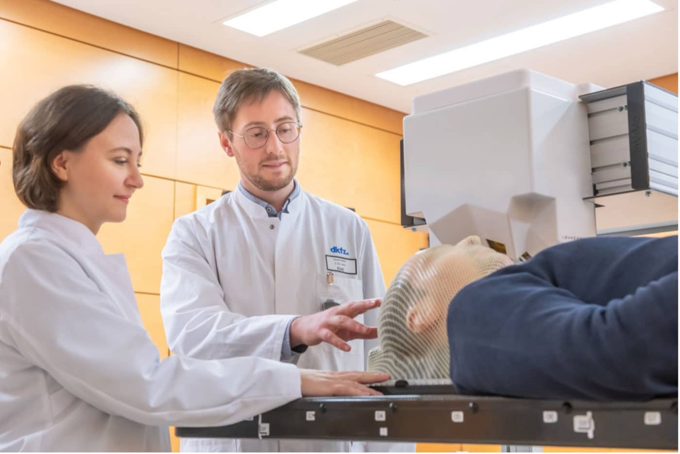
Mária Martišíková (left), the project leader from Heidelberg University Hospital and German Cancer Research Center (DKFZ), and DKFZ researcher Laurent Kelleter. Credit: Heidelberg University Hospital / H.Schroeder
Scientists are testing a new device that will help them more accurately target cancer cells during ion radiotherapy for head and neck tumors.
Scientists from the German National Center for Tumor Diseases (NCT), the German Cancer Research Center (DKFZ), and the Heidelberg Ion Beam Therapy Center (HIT) at Heidelberg University Hospital are currently testing the imaging device on their first patients. The device includes a small Timepix3 pixel detector developed at CERN, which allows head and neck tumors to be closely monitored during ion radiotherapy, making them easier to target and thus helping limit the treatment’s side effects.
“One of the most advanced methods for treating head and neck tumors involves irradiation with ion beams. This has one unique feature: it can be precisely tailored to the depth inside the human head where the particles should have the maximal effect”, explains Mária Martišíková, the head of the DKFZ team.
Like other forms of irradiation, ion radiation can have drawbacks. While it can be effective in targeting tumors, it can also affect healthy tissue surrounding the tumor. In the case of brain tumors, this can be particularly challenging, as damage to the optic nerve or a patient’s memory is possible.
Ideally, the irradiated area around the tumor should be minimized while maximizing the dose to the tumor. However, current technology does not yet allow for precise enough targeting of ions to achieve this ideal scenario.
It can be challenging to treat brain tumors, especially when the situation inside a patient’s head changes during therapy. Physicians typically use an X-ray computed tomography (CT) scan image taken before treatment as a “map” to target the tumor with ion beams.
However, the situation inside the skull can evolve during therapy, and there was no reliable tool to alert physicians in case of such changes. A new device called ADVACAM could help solve these issues by tracking the secondary particles created when ions pass through the head, improving the navigation of ion beams and ensuring more effective treatment.
“Our cameras can register every charged particle of secondary radiation emitted from the patient’s body. It’s like watching balls scattered by a billiard shot. If the balls bounce as expected according to the CT image, we can be sure we are targeting correctly. Otherwise, it’s clear that the ‘map’ no longer applies. Then it is necessary to replan the treatment,” describes Lukáš Marek from ADVACAM.
Using the camera to gather additional information can greatly enhance the effectiveness of the treatment. By analyzing the data obtained from the camera, it is possible to adjust the irradiation series in real time, which can lead to more accurate and effective treatment. The system can even correct the path of the ion beam in real time.
This is a great example of how technology developed for detectors used in fundamental physics research can be successfully transferred to the healthcare domain to benefit patients.
“When we started developing pixel detectors for the LHC, we had one target in mind: to detect and image each particle interaction and thereby help physicists unravel the secrets of nature at high energies. The Timepix detectors were developed by the multidisciplinary Medipix Collaborations, whose aim is to take the same technology to new fields. Many of those fields were completely unforeseen at the beginning, and this application is a brilliant example of that,” says Michael Campbell, spokesperson for the Medipix Collaborations.
Leave A Comment
You must be logged in to post a comment.