Source: www.laboratorytalk.com
Author: staff
A team of Indian cancer researchers led by Dr Narayanan Subhash has developed a simple, non-invasive spectral imaging system that holds the possibility of rapid, inexpensive mass screening. Even in the hands of non-clinical staff, it is capable of real-time discrimination of healthy oral tissue from pre-malignant and malignant tissues with accuracy comparable to the gold standard histopathology of a biopsy sample.
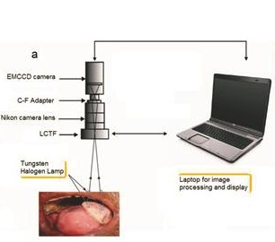
The core of the novel Diffuse Reflectance Imaging System (DRIS) is an Andor Luca-R EMCCD camera, which captures monochrome images of the patient’s mouth at 545 and 575 nm.
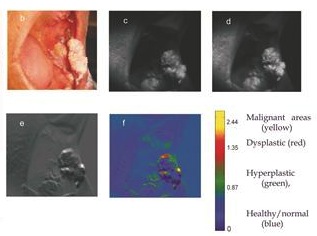
Andor’s SOLIS software computes a ratio image (R545/R575) of the area under investigation and generates a Pseudo Colour Map (PCM) where blue designates healthy tissue, red denotes dysplastic/pre-malignant tissue and yellow identifies malignant tissue.
This allows rapid visual differentiation of oral lesions and identification of regions with pre-malignant characteristics.
“Since mortality from oral cancer is particularly high, early detection, diagnosis and treatment is vital in increasing the survival rate of those with the disease,” says Dr Subhash. “Our imaging method has the great advantage of non-invasively scanning entire lesions and their surrounding areas and automatically categorising these oral lesions into normal/clinically healthy, pre-malignant, and malignant tissue in real-time.
“It also delineates the boundaries of neoplastic changes and locates sites with the most malignant potential for biopsy, thereby avoiding unnecessary repeated biopsies and delay in diagnosis. What’s more, imaging the entire region may also help the surgeons to identify the margins of the lesion that cannot be easily visualised by the naked eye during surgical interventions.”
Orla Hanrahan of Andor added: “The Luca-R EMCCD camera is well-equipped to handle this demanding role. It is built around a monochrome, megapixel frame transfer EMCCD sensor to deliver single photon detection sensitivity and unrestrained QE (65% max) in a TE cooled, USB 2.0 camera platform.”
Leave A Comment
You must be logged in to post a comment.