Source: www.medgadget.com
Author: staff
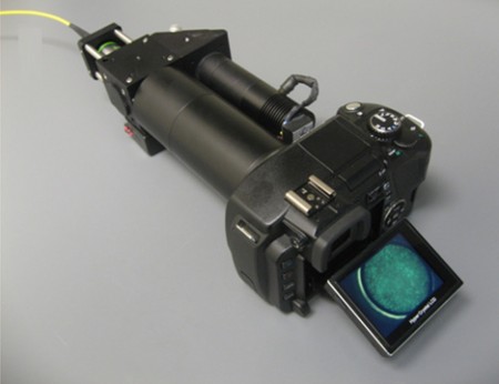
Photography-loving doctors now have more reasons to love their digital cameras. MacGyvers at Rice University and MD Anderson Cancer Center have cleverly engineered your everyday dSLR into a portable, high-resolution fiber-optic fluorescence imaging system that can detect cancer in-vivo.
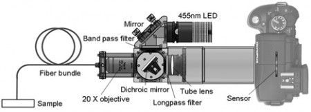
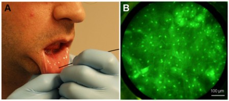
In this month’s PLoS ONE, they showed off the prowess of their camera system retrofitted with a LED light, an objective lens, a fiber-optic bundle in capturing sub-cellular images non-invasively and in real-time. In field tests of a fluorescence-labeled oral cancer cell culture, a surgically-resected human tissue specimen with dysplastic and cancerous regions, and a healthy human subject in vivo, the fiber-optic microscope resolved individual nuclei in all specimens and tissues imaged to distinguish qualitatively and quantitatively between normal, precancerous and/or cancerous tissues.
Portable and inexpensive at $2000 all-together, the clever device may be a useful tool to assist in the identification of early neoplastic changes in epithelial tissues in spartan clinical settings where MacGyver himself may have been.


Leave A Comment
You must be logged in to post a comment.