Source: medicalxpress.com
Author: University of Warwick
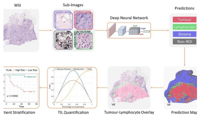
Whole slide images are multi-gigapixel images and cannot be used directly for image analysis tasks particularly training a deep learning based classifier. Therefore, we divide the WSIs into small regions (patches) for processing. A deep learning based classifier is applied on the patches to identify whether the patch contains tumour, lymphocytes or other histological primitives. However, the regions where the lymphocytes are infiltrating the tumour may not be confined within a patch. Besides, there is considerable variation in the size of TIL regions, making the quantification of TILs a non-trivial task. We address this issue by adopting the widely accepted definition of TILs, i.e., lymphocytes that lie in the neighbourhood of tumour areas. The patch labels predicted as lymphocytes or tumour are then used to compute a statistical measure of co-localization, which is further incorporated into the computation of the TILAb score of lymphocytic infiltration. Credit: University of Warwick
The chances of surviving oral cancers can be predicted by state of the art AI algorithms—developed by scientists at the Department of Computer Science at the University of Warwick—that precisely calculate the abundance of immune cells in the midst of tumour cells to help better understand the spread of and resistance to cancer.
In 2014 there were more than 11,000 cases of head and neck cancers in the UK and more than 2,300 deaths resulting from the most common of them; oral cavity cancer.
Oral cancer is most prevalent in South Asia, particularly India, Pakistan and Sri Lanka, mainly due to tobacco chewing, betel quid consumption and viral infections such as HPV (Human papillomavirus).
The chances of surviving such cancers can be predicted thanks to research in a pilot study from the University of Warwick’s Department of Computer Science in the paper “A Novel Digital Score for Abundance of Tumour Infiltrating Lymphocytes Predicts Disease Free Survival in Oral Squamous Cell Carcinoma,” published today, Monday 16 September, in the journal Nature Scientific Reports.
Researchers managed to develop a digital score that could measure Tumour Infiltrating Lymphocytes (TILs). The more TILs present the higher the chance of survival and longer disease free survival of oral cancer.
The images were produced by scans from patients at Shaukat Khanum Memorial Cancer Hospital Research Centre in Pakistan, who had all already been treated by radiation and a head and neck surgery. The cancer tissue samples were then sent to University Hospital Coventry and Warwickshire in the UK, where using a state of the art imaging machine researchers were able to digitally produce high-resolution images of the samples at a microscopic scale.
The presence of lymphocytes in the vicinity of the tumor cells doesn’t only help determine the stage of the cancer, it can be used to predict the progression of it accurately.
The more TILs present in the scans indicates the patient’s immunity to the cancer and the response to treatment, and the density and spatial arrangement of TILs correlates with the chances of overall survival and disease free survival.
Professor Nasir Rajpoot from the Department of Computer Science at the University of Warwick, who led the study, comments:
“We are only beginning to unravel the remarkable potential of wealth of information present in pathology image data. This pilot study shows that with the help of modern cancer image analytics algorithms, we can precisely calculate the score of abundance of TILs in oral cancers in an objective manner and then use that score for risk stratification in terms of disease free survival.”
Prof Hisham Mehanna, Professor of Head & Neck Oncology at the University of Birmingham, comments:
“This is a very exciting development. Not only is this one of the first artificial intelligence based scores to be validated in oral cancer, this score also seems to have a strong prognostic power, which could eventually lead to stratifying patients for different treatment modalities.”
Dr. Asif Loya, Medical Director at the Shaukat Khanum Memorial Cancer Hospital & Research Centre in Pakistan where the patient samples were sourced from, comments:
“With almost 13,000 new cases every year, oral cancers have the highest incidence rates among cancers in Pakistan, second highest mortality rate, and a very low five-year survival. However, there is little known about the histological signatures corresponding to patient subgroups with differing outcomes in this part of the world. Histologic risk assessment is strongly predictive of local disease-free and overall survival in oral squamous cell carcinoma thus there is need for a validated scoring system to be used as an aid in treatment decision making in these cancers in our patients . Strong pilot data from this collaborative research using the objective assessment method of digital analysis may play a role in establishing such prognostication models so that treatment decisions related to elective neck dissection (END) and adjuvant radiotherapy can be made more appropriately.”
Leave A Comment
You must be logged in to post a comment.